Diagnosis of Health

Wednesday 1 June 2011

Hepatocellular carcinoma better depicted with contrast CT than with ultrasound.

HISTORY: Right upper quadrant pain and elevated liver function tests.


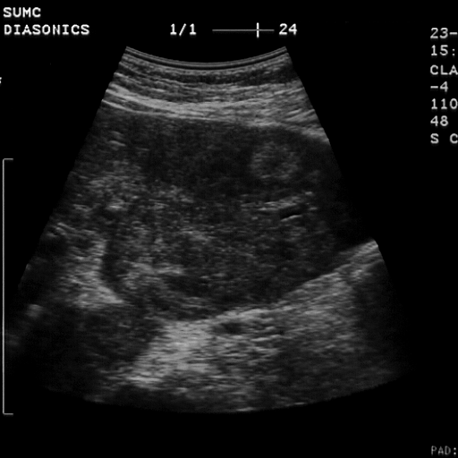
FINDINGS: Images 1-3 are scans of the right lobe of the liver
demonstrating marked heterogeneity of the hepatic parenchymal
echogenicity.  Subtle refractive shadowing is noted in Image 1.  In
Image 2 there is evidence of a vague echogenic mass seen centrally in
the right lobe of the liver.

Images 4-6 are contrast-enhanced CTs demonstrating cirrhosis with a
nodular liver contour.  There is perihepatic ascites and a large right
lobe liver mass.  On Image 5 there is evidence of portal venous
invasion.

DIAGNOSIS: Hepatocellular carcinoma better depicted with contrast CT
than with ultrasound.

DISCUSSION: In patients with advanced cirrhosis, the sonographic
diagnosis of hepatocellular carcinoma may be quite challenging.  Fatty
infiltration, regenerative nodules, and confluent fibrosis all degrade
the sonographic evaluation of the liver.  Nevertheless, refractive
shadows are very worrisome for a space occupying mass as evident in
Image 1 of this case.  CT more clearly depicted the parenchymal mass and
clearly demonstrated the portal venous invasion to better advantage.


Calcified liver metastasis secondary to prostatic carcinoma.

HISTORY: Prostate carcinoma.


FINDINGS: Images 1-4 are transverse scans of the right lobe of the liver.  Note the large right pleural effusion and the discrete mass seen on Image 5 displacing the middle and left hepatic veins.  Note in Image 6 that there appears to be shadowing from an echogenic portion of the mass consistent with calcification.
 
DIAGNOSIS: Calcified liver metastasis secondary to prostatic carcinoma.

DISCUSSION: Calcification is unusual in most hepatic metastasis.
Differential diagnosis includes metastatic osteogenic sarcoma, mutinous adenocarcinoma and, as in this case, prostate carcinoma.  Calcification may occur in primary tumors such as hepatocellular carcinoma, cavernous hemangioma, and in benign lesions such as an old abscess or hematoma.
The hypoechoic peripheral halo around the lesion in this case is typical for metastatic disease.


Benign hemorrhagic hepatic cyst.

HISTORY: 73-year-old female with right upper quadrant pain.


FINDINGS: Image 1 is a color Doppler sonogram of the right lobe of the liver demonstrating a large complex cystic lesion that contains tissue along the inner aspect of the cyst wall.  Notice that the "internal
tissue" is entirely avascular with color Doppler sonography.
 
Noncontrast CT scans demonstrate a large nondescript cystic lesion.

DIAGNOSIS: Benign hemorrhagic hepatic cyst.

DISCUSSION: The differential diagnosis of this complex cystic lesion includes abscess, hematoma, biloma, cystic neoplasm and, as in this
case, a hemorrhagic cyst.  Parasitic cysts may also be included in the
differential diagnosis.  However, there are often more specific
features, such as daughter cysts or calcification of the cyst wall, that
are not present in this case.  Amoebic abscesses may be perfectly round or oval in configuration and demonstrate no evidence of calcification.

The failure of color Doppler to show any intrinsic vascularity within
the mural tissue on ultrasound was an important observation.  At
pathologic examination this proved to be an area of clot within the
hemorrhagic cyst.  Clot, of course, would be avascular unlike the mural nodules of malignant lesions such as biliary cyst adenocarcinoma or
metastatic ovarian carcinoma.


Gastric carcinoma with extensive liver metastasis.

 
HISTORY: Early satiety and heme positive stool.  Elevated liver function
tests.  Rule out biliary obstruction.


FINDINGS: Images 1-6 are sagittal and transverse scans of the stomach
and left lobe of the liver.  In Images 1 and 2 there is asymmetric
thickening of the body of the stomach.  Multiple liver metastasis are
evident.  Note that some of the lesions are predominantly hypoechoic
(Image 6) while other lesions are echogenic with peripheral halos (Image
4).

DIAGNOSIS: Gastric carcinoma with extensive liver metastasis.
 
DISCUSSION: This case nicely illustrates the variable appearance of
hepatic metastasis.  Both hypo- and hyperechogenic lesions are
demonstrated.  Peripheral hypoechoic halos surrounding focal echogenic
lesions are typical features of malignant disease.  The demonstration of
asymmetric thickening of the gastric wall was fortuitous in this case.
However, this indeed proved to be the cause of the patient's metastasis
as gastric carcinoma was discovered at endoscopy.