HISTORY: Prostate carcinoma.
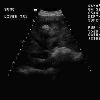
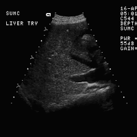
 FINDINGS: Images 1-4 are transverse scans of the right lobe of the liver. Note the large right pleural effusion and the discrete mass seen on Image 5 displacing the middle and left hepatic veins. Note in Image 6 that there appears to be shadowing from an echogenic portion of the mass consistent with calcification.
DIAGNOSIS: Calcified liver metastasis secondary to prostatic carcinoma.
DISCUSSION: Calcification is unusual in most hepatic metastasis.
 Differential diagnosis includes metastatic osteogenic sarcoma, mutinous adenocarcinoma and, as in this case, prostate carcinoma. Calcification may occur in primary tumors such as hepatocellular carcinoma, cavernous hemangioma, and in benign lesions such as an old abscess or hematoma.
The hypoechoic peripheral halo around the lesion in this case is typical for metastatic disease.


No comments:
Post a Comment