HISTORY: Right upper quadrant pain and elevated liver function tests.
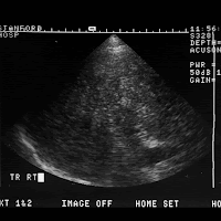
FINDINGS: Images 1-3 are scans of the right lobe of the liver
demonstrating marked heterogeneity of the hepatic parenchymal
Image 2 there is evidence of a vague echogenic mass seen centrally in
the right lobe of the liver.
Images 4-6 are contrast-enhanced CTs demonstrating cirrhosis with a
lobe liver mass. On Image 5 there is evidence of portal venous
invasion.
DIAGNOSIS: Hepatocellular carcinoma better depicted with contrast CT
DISCUSSION: In patients with advanced cirrhosis, the sonographic
diagnosis of hepatocellular carcinoma may be quite challenging. Fatty
infiltration, regenerative nodules, and confluent fibrosis all degrade
shadows are very worrisome for a space occupying mass as evident in
Image 1 of this case. CT more clearly depicted the parenchymal mass and
clearly demonstrated the portal venous invasion to better advantage.


No comments:
Post a Comment