

HISTORY: Pancreatic tail mass on outside CT scan.
FINDINGS: Images 1-3 are transverse scans of the left lobe of the liver
 demonstrating diffuse heterogeneity and alteration in the normal
parenchymal echogenicity. Notice in Image 1 there is bulging of the
contour of the liver due to an echogenic mass.
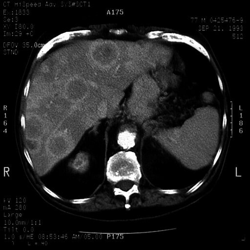
Images 4, 5, and 6 are a contrast-enhanced CT scans of the liver and
pancreas demonstrating multiple hepatic lesions and a calcified mass in
the tail of the pancreas.
DIAGNOSIS: Multifocal hepatoma. Calcified neuroendocrine tumor of the
pancreas.
DISCUSSION: This case illustrates the fact that well defined areas of

contrast CT, note the peripheral enhancing rim around the left lobe
 lesions. The calcified pancreatic mass was not well imaged with
sonography due to its location in the tail in the pancreas.
Calcification is rarely seen in ductal adenocarcinomas of the focal
hepatic lesions. Differential diagnosis includes multifocal hepatoma,
metastatic carcinoma, and neuroendocrine tumor.
No comments:
Post a Comment