HISTORY: A 56-year-old male with known hepatitis B. Weight loss and
right upper quadrant pain.
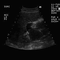
FINDINGS: Images 1 and 2 are transverse and longitudinal images of the
right lobe of the liver demonstrating a complex solid mass (arrows,
enlargement of the portal vein by a diffuse hypoechoic mass (arrows,
Image 3). Note on Image 4, which is a longitudinal scan of the left
portal vein, there is a thrombus extending to the proximal portion of
the anterior division of the left portal vein.
Image 5 is a B- color image of the right lower lobe of the liver
demonstrating branching hypoechoic tumor thrombus within the anterior
posterior divisions of the right portal vein. Image 6 is a color
flow within the tumor thrombus. Image 7 is a spectral Doppler tracing
documenting arterial flow within the portal vein. Image 8 is a
transverse scan of the portahepatis demonstrating numerous periportal
color Doppler scan of the right lobe of the liver demonstrating the
large mass which is predominantly avascular.
DIAGNOSIS: Hepatocellular carcinoma with portal vein invasion.
DISCUSSION: The sonographic findings in this case clearly demonstrate
evidence of tumor thrombus within the portal vein from the patient's
hepatocellular carcinoma. The color Doppler demonstration of intrinsic
tumor invasion rather than bland thrombus. Although the tumor mass
within the right lobe is relatively avascular, this is somewhat unusual
for primary hepatocellular carcinoma. In 75% of patients, significant
sonography. In only 25% of cases the lesions will appear hypo- or
avascular. Portal venous invasion, although typical for hepatocellular
carcinoma, may also occur in cholangiocarcinoma or metastases.
Percutaneous biopsy of the portal vein thrombus may also be helpful to
distinguish bland thrombus from neoplastic invasion.


No comments:
Post a Comment