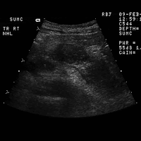
 HISTORY: Known non-Hodgkin's lymphoma with left upper quadrant pain.
FINDINGS: Images 1-5 are longitudinal and transverse scans of the upper
abdomen including images of the liver, pancreas, and kidney. Images 1
and 2 are transverse scans of the liver demonstrating diffuse hypoechoic
infiltration of the left hepatic lobe. Note intrahepatic bile duct
dilatation within the medial segment of the left lobe. Image 3 is a
(arrows) involving the body and tail of the pancreas. Images 4 and 5
are sagittal scans of the left kidney demonstrating complete loss of
architecture of the left kidney with hypoechoic masses replacing the
Images 6-8 are contrast-enhanced CT scans of the upper abdomen
confirming multiple low density masses involving the liver, pancreas,
and left kidney.
 DIAGNOSIS: Non-Hodgkin's lymphoma involving the liver, kidney, and
pancreas.
DISCUSSION: Abdominal lymphoma is typically hypoechoic with sonography.
The multicentric involvement in this case is unusual given the relative
paucity of nodal disease. The visceral involvement is much more
commonly encountered with non-Hodgkin's lymphoma. AIDS-related
lymphoma will frequently demonstrate this degree of visceral involvement
in the absence of large bulky nodal disease. Included in the differential
diagnosis are metastatic adenocarcinoma, melanoma, and sarcoma


No comments:
Post a Comment