
HISTORY: 21-year-old female with known orbital sarcoma, now with
elevated liver function tests.

FINDINGS: Images 1-3 are transverse scans of the liver demonstrating
 multiple rounded echogenic masses (arrows) with marked compression of
the portal venous system as seen on Image 3.
Images 4 and 5 are contrast-enhanced spiral CT scans demonstrating
 extensive necrosis within both lesions as there is central liquefaction.
DIAGNOSIS: Metastatic sarcoma to the liver.
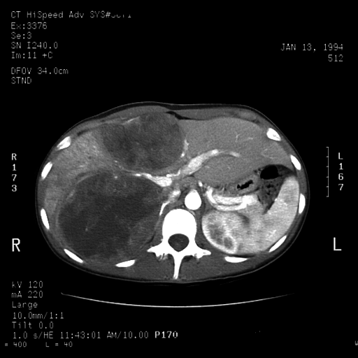 DISCUSSION: In this patient the right lobe of the liver is virtually
replaced by metastatic lesions. There is marked compression of the
right portal vein. Because of a lack of a hepatic contrast agent to
assess parenchymal perfusion, ultrasound is relatively limited in the
diagnosis of tumor necrosis. On contrast-enhanced CT, note that there
is viable tumor on the periphery of lesions, and central liquefaction
and necrosis. This is of relevance in performing percutaneous liver
biopsies as only blood and necrotic tissue will be obtained if the
center of the lesion is biopsied.
No comments:
Post a Comment