HISTORY: Rectal carcinoma. Intraoperative evaluation of liver mets.

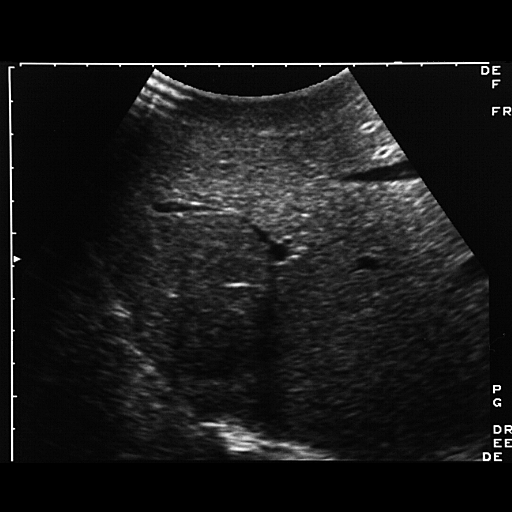


FINDINGS: Images 1-4 are intraoperative sonograms of the liver to
evaluate for metastases. Images 1-3 demonstrate a 3 x 5 cm hypoechoic
mass seen abutting the diaphragm in the junction of the anterior and
posterior segments of the right lobe (arrows). Note the right hepatic
vein in Images 2 and 3. On Image 11 there is clear evidence of
refractive edge shadowing from the lesion. On Image 4 there is a very
subtle hypoechoic lesion which was an additional satellite nodule seen
high up near the diaphragm (arrows). Surgical resection of this lesion
confirmed an additional metastatic deposit.
DIAGNOSIS: Hepatic metastases from rectal carcinoma.
DISCUSSION: Intraoperative ultrasound has two main advantages: 1) it
improves sensitivity for detection of lesions, and 2) it provides useful
information regarding the relationship of metastases to major hepatic
vessels. Lesions that are located in close proximity to the confluence
of the hepatic veins are often non-resectable. In selected cases,
sonography may detect up to 16% more lesions than are not clinically
palpable at the time of surgery.
No comments:
Post a Comment