HISTORY: Rising LFF's.
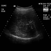
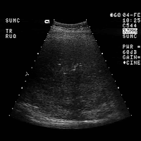
notice there is a clear cut area of refractive shadowing (arrowheads)
seen adjacent to a focal echogenic lesion. Images 2-5 demonstrate fine
punctate areas of increased echogenicity consistent with calcifications
(arrows). Notice the hypoechoic halo around the inferior margin of the
lesion seen on Image 5. Image 6 is a transverse scan of the left lobe
of the liver at the level of the celiac axis. Notice the celiac axis
adenopathy.
DIAGNOSIS: Metastatic mucinous adenocarcinoma of the colon.
DISCUSSION: Calcification may occur in a variety of hepatic metastases,
but it is most commonly encountered with mucinous adenocarcinoma. Note
the typical sonographic features of solid masses of the liver with
refractive shadows and hypoechoic halos. Regional lymphadenopathy canbe also noted with a wide variety of metastatic diseases, but given the
punctate areas of calcifications metastatic colon cancer is the most
likely diagnostic possibility.


No comments:
Post a Comment