HISTORY: Vague right upper quadrant pain with a prior history of lung cancer.
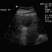
FINDINGS: Images 1 through 3 demonstrate multiple hypoechoic masses
within the right lobe of the liver (arrows). Note on Images 3 and 4
there is slight enhanced through sound transmission.
DISCUSSION: There is a broad spectrum of the sonographic appearance of
hepatic metastasis. The lesions run the entire gamut from predominantly
metastasis (as in this case) or echogenic lesions (as seen in both
metastatic colon and breast carcinoma). Not infrequently there is a
peripheral hypoechoic halo around the lesion due to rapid enlargement of
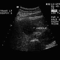
the lesion or surrounding compressed hepatic parenchyma. A biopsy was
performed under ultrasound guidance (Image 4). Note the needle
placement within the lesion. These findings are compatible with
squamous cell carcinoma from the patient's known lung cancer.
Biopsy is often essential to accurately diagnose the precise cause of a
liver mass. In addition to metastatic disease, abscesses and other
complex fluid collections must be included in the differential
diagnosis.


No comments:
Post a Comment