HISTORY: 59-year-old male with hepatitis-B; rule out hepatoma.
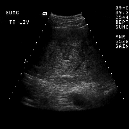
 FINDINGS: Images 1 and 2 are sagittal and transverse scans of the left
lobe of the liver demonstrating a focal hepatic mass that is
characterized by peripheral hypoechogenicity and central areas of
increased echogenicity. In addition, Image 3 demonstrates a small
echogenic focus in the right lobe.
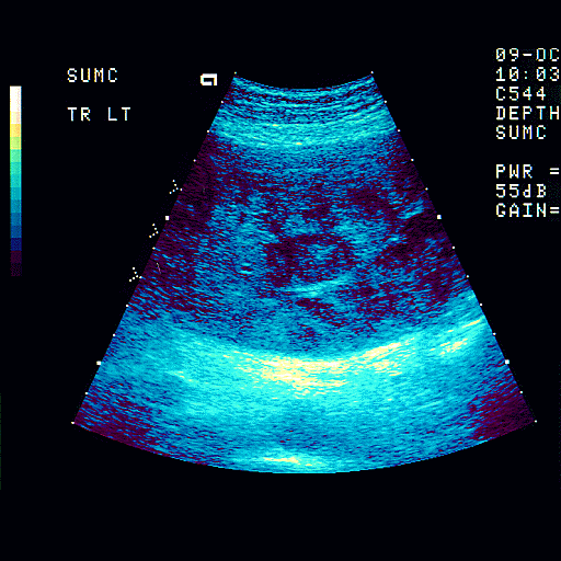
 Image 4 is a B-color image demonstrating a "mosaic" pattern of the left
lobe mass. Color Doppler Images 5-7 demonstrate increased flow within
the left lobe mass.
DIAGNOSIS: Left lobe hepatoma in a patient with known hepatitis-B.
DISCUSSION: The sonographic features of hepatocellular carcinoma are
variable depending upon parenchymal and/or vascular invasion. Lesions
may be hypoechoic, echogenic, or of mixed echogenicity. Several
 different morphologic features have been described including nodular or
conglomerate patterns and/or infiltrative patterns. Not infrequently
hepatocellular carcinoma is multicentric, as in this case where there is
a small echogenic nidus of tumor noted in the right lobe. The
 sonographic pattern in this case may be described as a "mosaic pattern"
due to the alternating pattern of echogenic rounded foci and sonolucent
peripheral halos. It is important to interrogate the portovenous system
with color Doppler to detect venous invasion in any patient with a focal
parenchymal mass. Venous invasion strongly suggests primary
hepatocellular carcinoma. With color Doppler, hepatocellular carcinoma
has intrinsic vasculature approximately 75% of the time. Alternatively
patients with metastasis to the liver have extrinsic vascularity along
their contour 75% of the time and lack internal intrinsic vascularity.
No comments:
Post a Comment