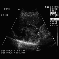
HISTORY: 64-year-old female with known ovarian carcinoma.
FINDINGS: Images 1-3 are longitudinal and transverse scans of the liver.
Image 4 is a sagittal scan of the spleen. Note that Images 1-3
demonstrate a complex cystic mass along the edge of the liver surface.
It contains irregular septations and mural nodules. Image 1
demonstrates that the mass extends exophytically from the liver surface.
Image 4 is a sagittal scan of the spleen demonstrating a hypoechoic, but
predominantly solid lesion near the diaphragm.
DIAGNOSIS: Ovarian carcinoma with cystic metastases to the liver and
spleen.
DISCUSSION: Ovarian carcinoma may produce cystic metastases in the
liver, spleen, peritoneal cavity, and omentum. The appearance of the
hepatic lesion is not unlike the appearance of the primary tumor,
demonstrating a complex cystic mass with mural nodules and irregularly
thickened septations. When the lesions are located along the peripheral
margin of the liver, it is often difficult to know whether the lesion
originates within the hepatic parenchyma or is located along the
peritoneal surface of the liver with simple mass effect on the liver.


No comments:
Post a Comment