Diagnosis of Health

Wednesday, 1 June 2011

Hepatocellular carcinoma invading the portal vein.

HISTORY: Vague right upper quadrant pain with elevated liver function
tests.


FINDINGS: Images 1-6 are transverse and longitudinal scans of the liver.
Note there is a heterogeneity of the hepatic parenchymal echogenicity in
Images 1-4.  In Image 4 there appears to be a more discrete echogenic
mass within the liver that casts some refractive shadows.  Image 5 is a
sagittal scan of the main portal vein demonstrating diffuse level echoes
within the portal vein and distention of the walls of the vein.  Image 6
is a color Doppler sonogram demonstrating no flow within the portal vein
with a patent hepatic artery.

Images 7 and 8 are CT scans of the liver obtained with intravenous
contrast enhancement.  Notice the large infiltrating mass replacing much
of the right lobe of the liver, and in Image 8 an invasion of the main
portal vein.  Only the hepatic artery is visible in the porta hepatis.

DIAGNOSIS: Hepatocellular carcinoma invading the portal vein.

DISCUSSION: In patients with infiltrating neoplasms such as
hepatocellular carcinoma, CT can better delineate the extent of hepatic
parenchymal tumor.  On the other hand, color Doppler sonography is often
a very easy technique to confirm the presence of portal vein thrombosis.
In selected patients, arterial flow can actually be identified within
the viable tumor thrombus.


Monday, 30 May 2011

Focal nodular hyperplasia associated with cavernous hemangioma of the liver.


HISTORY: Right upper quadrant pain, rule out gallstones.


FINDINGS: Images 1-5 are transverse scans of the liver.  In Image 1
there is a well-defined echogenic mass seen in the posterior segment of
the right lobe of the liver.  Images 2 and 3 demonstrate a mass in the
anterior segment of the right lobe that has slightly increased
echogenicity compared to normal liver (arrows, Image 2).  Images 4 and 5
are color Doppler sonograms of the mass in the anterior segment of the
right lobe demonstrating intrinsic flow.

DIAGNOSIS: Focal nodular hyperplasia associated with cavernous
hemangioma of the liver.
 
DISCUSSION: There is an increased incidence of focal nodular hyperplasia
(FNH) in patients with hemangioma of the liver.  Sonographically,
hemangiomas demonstrate a variable appearance, but when small, they are
typically hyperechoic in appearance with slight distal enhanced through
sound transmission.  An important negative finding is lack of a
peripheral hypoechoic halo and a lack of refractive shadowing.  FNH may
be quite subtle to detect with grayscale imaging as the echogenicity is
very similar to the normal liver.  The color Doppler sonograms are
useful to demonstrate increased intrinsic flow within the lesion quite
typical of FNH.  Intrinsic flow can also be seen with malignant lesions
such as hepatocellular carcinoma and some metastatic lesions.

Therefore, this finding is nonspecific and requires other confirmatory
studies such as nuclear medicine examination or MRI.


Cirrhosis, portal hypertension, and hepatocellular carcinoma.


HISTORY: Rising liver function test protein in a patient with known
cirrhosis.


FINDINGS: Images 1-3 are high-resolution linear scans of the left lobe
of the liver demonstrating a well-defined echogenic mass.  In Image 2,
there is clear-cut evidence of distal acoustic shadowing from the mass.
The liver capsule is bowed by the lesion.  Image 3 is a pulse Doppler
sonogram demonstrating high velocity vessels within the lesion.

Contrast-enhanced CT images demonstrate a recanalized umbilical vein
(best seen on Images 4 and 5) as well as a small focal lesion seen on
the lateral segment of the left lobe corresponding to the ultrasound
abnormalities.  Note also the perisplenic varices on Image 7.

Percutaneous FNA biopsy revealed hepatocellular carcinoma.

DIAGNOSIS: Cirrhosis, portal hypertension, and hepatocellular carcinoma.


DISCUSSION: Small hepatocellular carcinomas may be quite echogenic and
may mimic hemangiomas in a patient with know cirrhosis.  Unlike
hemangiomas, they may cause distal acoustic shadowing as well as have
intrinsic high velocity arterial flow.  Most hemangiomas are avascular
with color Doppler sonography and if anything cause slight distal
enhanced through sound transmission due to the fluid nature of the mass
(just tangled collections of hepatic sinusoids).


Neuroendocrine tumor of the pancreas.


HISTORY: Pancreatic tail mass on outside CT scan.


FINDINGS: Images 1-3 are transverse scans of the left lobe of the liver
demonstrating diffuse heterogeneity and alteration in the normal
parenchymal echogenicity.  Notice in Image 1 there is bulging of the
contour of the liver due to an echogenic mass.

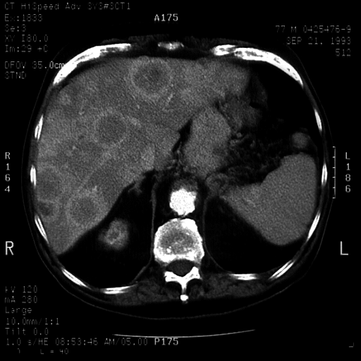
Images 4, 5, and 6 are a contrast-enhanced CT scans of the liver and
pancreas demonstrating multiple hepatic lesions and a calcified mass in
the tail of the pancreas.
 
DIAGNOSIS: Multifocal hepatoma.  Calcified neuroendocrine tumor of the
pancreas.

DISCUSSION: This case illustrates the fact that well defined areas of
tumor on contrast CT may appear quite ill defined on ultrasound.  On
contrast CT, note the peripheral enhancing rim around the left lobe
lesions.  The calcified pancreatic mass was not well imaged with
sonography due to its location in the tail in the pancreas.
Calcification is rarely seen in ductal adenocarcinomas of the focal
hepatic lesions.  Differential diagnosis includes multifocal hepatoma,
metastatic carcinoma, and neuroendocrine tumor.