Diagnosis of Health

Monday, 30 May 2011

Focal nodular hyperplasia associated with cavernous hemangioma of the liver.


HISTORY: Right upper quadrant pain, rule out gallstones.


FINDINGS: Images 1-5 are transverse scans of the liver.  In Image 1
there is a well-defined echogenic mass seen in the posterior segment of
the right lobe of the liver.  Images 2 and 3 demonstrate a mass in the
anterior segment of the right lobe that has slightly increased
echogenicity compared to normal liver (arrows, Image 2).  Images 4 and 5
are color Doppler sonograms of the mass in the anterior segment of the
right lobe demonstrating intrinsic flow.

DIAGNOSIS: Focal nodular hyperplasia associated with cavernous
hemangioma of the liver.
 
DISCUSSION: There is an increased incidence of focal nodular hyperplasia
(FNH) in patients with hemangioma of the liver.  Sonographically,
hemangiomas demonstrate a variable appearance, but when small, they are
typically hyperechoic in appearance with slight distal enhanced through
sound transmission.  An important negative finding is lack of a
peripheral hypoechoic halo and a lack of refractive shadowing.  FNH may
be quite subtle to detect with grayscale imaging as the echogenicity is
very similar to the normal liver.  The color Doppler sonograms are
useful to demonstrate increased intrinsic flow within the lesion quite
typical of FNH.  Intrinsic flow can also be seen with malignant lesions
such as hepatocellular carcinoma and some metastatic lesions.

Therefore, this finding is nonspecific and requires other confirmatory
studies such as nuclear medicine examination or MRI.


Cirrhosis, portal hypertension, and hepatocellular carcinoma.


HISTORY: Rising liver function test protein in a patient with known
cirrhosis.


FINDINGS: Images 1-3 are high-resolution linear scans of the left lobe
of the liver demonstrating a well-defined echogenic mass.  In Image 2,
there is clear-cut evidence of distal acoustic shadowing from the mass.
The liver capsule is bowed by the lesion.  Image 3 is a pulse Doppler
sonogram demonstrating high velocity vessels within the lesion.

Contrast-enhanced CT images demonstrate a recanalized umbilical vein
(best seen on Images 4 and 5) as well as a small focal lesion seen on
the lateral segment of the left lobe corresponding to the ultrasound
abnormalities.  Note also the perisplenic varices on Image 7.

Percutaneous FNA biopsy revealed hepatocellular carcinoma.

DIAGNOSIS: Cirrhosis, portal hypertension, and hepatocellular carcinoma.


DISCUSSION: Small hepatocellular carcinomas may be quite echogenic and
may mimic hemangiomas in a patient with know cirrhosis.  Unlike
hemangiomas, they may cause distal acoustic shadowing as well as have
intrinsic high velocity arterial flow.  Most hemangiomas are avascular
with color Doppler sonography and if anything cause slight distal
enhanced through sound transmission due to the fluid nature of the mass
(just tangled collections of hepatic sinusoids).


Neuroendocrine tumor of the pancreas.


HISTORY: Pancreatic tail mass on outside CT scan.


FINDINGS: Images 1-3 are transverse scans of the left lobe of the liver
demonstrating diffuse heterogeneity and alteration in the normal
parenchymal echogenicity.  Notice in Image 1 there is bulging of the
contour of the liver due to an echogenic mass.

Images 4, 5, and 6 are a contrast-enhanced CT scans of the liver and
pancreas demonstrating multiple hepatic lesions and a calcified mass in
the tail of the pancreas.
 
DIAGNOSIS: Multifocal hepatoma.  Calcified neuroendocrine tumor of the
pancreas.

DISCUSSION: This case illustrates the fact that well defined areas of
tumor on contrast CT may appear quite ill defined on ultrasound.  On
contrast CT, note the peripheral enhancing rim around the left lobe
lesions.  The calcified pancreatic mass was not well imaged with
sonography due to its location in the tail in the pancreas.
Calcification is rarely seen in ductal adenocarcinomas of the focal
hepatic lesions.  Differential diagnosis includes multifocal hepatoma,
metastatic carcinoma, and neuroendocrine tumor.


Non-Hodgkin's lymphoma

HISTORY: Known non-Hodgkin's lymphoma with left upper quadrant pain.


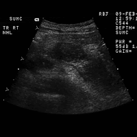
FINDINGS: Images 1-5 are longitudinal and transverse scans of the upper
abdomen including images of the liver, pancreas, and kidney.  Images 1
and 2 are transverse scans of the liver demonstrating diffuse hypoechoic
infiltration of the left hepatic lobe.  Note intrahepatic bile duct
dilatation within the medial segment of the left lobe.  Image 3 is a
transverse scan of the pancreas demonstrating multiple hypoechoic masses
(arrows) involving the body and tail of the pancreas.  Images 4 and 5
are sagittal scans of the left kidney demonstrating complete loss of
architecture of the left kidney with hypoechoic masses replacing the
parenchyma.

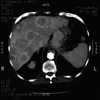
Images 6-8 are contrast-enhanced CT scans of the upper abdomen
confirming multiple low density masses involving the liver, pancreas,
and left kidney.

DIAGNOSIS: Non-Hodgkin's lymphoma involving the liver, kidney, and
pancreas.

DISCUSSION: Abdominal lymphoma is typically hypoechoic with sonography.
The multicentric involvement in this case is unusual given the relative
paucity of nodal disease.  The visceral involvement is much more
commonly encountered with non-Hodgkin's lymphoma.  AIDS-related
lymphoma will frequently demonstrate this degree of visceral involvement
in the absence of large bulky nodal disease.  Included in the differential
diagnosis are metastatic adenocarcinoma, melanoma, and sarcoma


Pleural metastases from carcinoma of the breast.

HISTORY: Right upper quadrant pain in a 62-year-old female with known breast carcinoma.

FINDINGS: Images 1-4 are intercostal scans of the dome of the liver and
right pleural space obtained with the patient in an upright position.
Note that there is hypoechoic pleural fluid as well as echogenic tissue
(arrow, Image 1) within the pleural space.  Image 4 is a spectral
Doppler tracing demonstrating arterial flow from the echogenic pleural
mass.
 
DIAGNOSIS: Pleural metastases from carcinoma of the breast.

DISCUSSION: Pleuritic pain (as in this patient) may at times be
clinically mistaken for abdominal pathology such as acute cholecystitis,
liver abscess, etc.  The echogenic pleural based tissue demonstrated
arterial flow on color Doppler.  This finding strongly suggested pleural
based metastases.  The differential diagnosis includes primary pleural
tumor such as mesothelioma as well as metastases.  Hemothorax or empyema
within the pleural space is avascular with color Doppler.